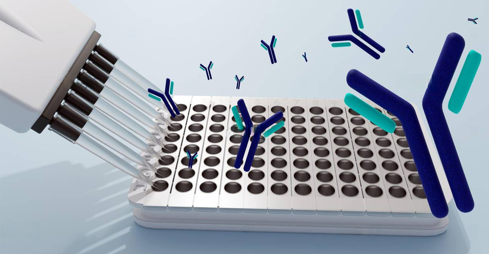
Structure of Antibodies
Antibodies, or immunoglobulins (Ig), are complex glycoproteins synthesized by B lymphocytes, playing a pivotal role in adaptive immunity. Their primary function is to identify and neutralize pathogens through specific antigen recognition.
Structural Components:
Heavy and Light Chains: Each antibody consists of two identical heavy chains (~50 kDa each) and two identical light chains (~25 kDa each), linked by disulfide bonds, forming a heterotetrameric structure.
Variable (V) and Constant (C) Regions:
Variable Regions: Located at the N-termini of both heavy and light chains, these regions exhibit significant amino acid diversity, enabling the binding to a vast array of antigens.
Constant Regions: These regions determine the antibody’s isotype and mediate effector functions by interacting with various immune components.
Wikipedia Complementarity-Determining Regions (CDRs): Within the variable regions, three hypervariable loops (CDR1, CDR2, CDR3) on each chain form the antigen-binding site. The diversity in these loops is central to the antibody’s specificity.
Nature Framework Regions (FRs): Interspersed between CDRs, FRs provide a scaffold that maintains the structural integrity of the variable regions. Mutations in FRs can influence the orientation and conformation of CDRs, thereby affecting antigen binding affinity.


Classes of Antibodies
IgG: Predominant in serum, capable of crossing the placenta to confer passive immunity to the fetus.
IgA: Abundant in mucosal secretions, serving as the first line of defense against inhaled or ingested pathogens.
IgM: Forms pentamers, facilitating efficient agglutination and initial immune responses.
IgE: Binds to mast cells and basophils, playing a key role in allergic reactions and defense against parasitic infections.
IgD: Functions primarily as a receptor on naive B cells, though its exact role remains less defined.
Monoclonal vs. Polyclonal Antibodies
Antibodies are classified based on their source and production methods. The two main types used in research and therapeutic applications are monoclonal antibodies (mAbs) and polyclonal antibodies (pAbs). The choice between them depends on the application, required specificity, and tolerance for variability.
Definition:
Monoclonal antibodies are derived from a single B lymphocyte clone and are thus homogenous in nature. They recognize one specific epitope on the target antigen. Production is typically achieved via hybridoma technology, wherein a B cell producing the desired antibody is fused with an immortalized myeloma cell, forming a hybrid cell line capable of continuous, monoclonal antibody production.
Modern Advances:
- Recombinant Monoclonal Antibodies (r-mAbs): Generated using transfected cell lines (e.g., CHO, HEK293), these provide animal-free production and greater control over isotype, Fc region, and glycosylation.
- Phage Display & Single B Cell Cloning: These allow for epitope mapping, humanization, and affinity maturation.
Advances:
- High Specificity & Low Background: Targets a single epitope, minimizing non-specific interactions.
- Reproducibility: Batch-to-batch consistency critical for diagnostics, drug development, and GMP workflows.
- Epitope Mapping: Useful for structural biology, mechanistic studies, or therapeutic blocking.
Limitations:
- Epitope Conformational Dependence: Some monoclonals may not recognize denatured proteins (e.g., in WB vs. native IHC).
- Time- and Resource-Intensive Production: From hybridoma screening to stability testing.
- Potential Loss of Reactivity: If antigen undergoes mutation or structural changes, the mAb may fail to bind.
Polyclonal Antibodies (pAbs)