Protocols

Multicolor Flow Cytometry Protocols

During flow cytometry, multicolor antibody staining is the common requirement. Some multicolor flow cytometry protocols should be followed during color matching.

1. Strongly/Weakly Expressed Antigens

Weakly expressed antigens match with strong fluorochromes. Strongly expressed antigens match with weak fluorochromes. This matching protocol helps to improve signal-to-noise ratio. Then, the signal of weakly expressed antigens will not be covered by strongly expressed antigens.

Applications: During antibody selection, suitable fluorochromes depend on antigen expression. E.g. for strongly expressed antigen CD45, weaker fluorochrome like FITC is proper. For weakly expressed antigens(e.g. some cytokine receptors), stronger fluorochrome like APC may be required.

2. Avoid Fluorescent Leak

Co-expressed antigens: For antigens co-expressed on the same cell, avoid to use interfered fluorochromes. Fluorochrome combinations with less spectral overlap are required. In the figure below, CD25 and Foxp3 antigens are co-expressed antigens.

Fluorescent Leak

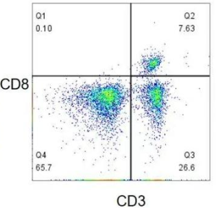
Mutually exclusive antigens refer to antigens won’t be co-expressed on the same cell. In the figure below, CD4 and CD8 are not co-expressed on the same cell, allowing slight fluorescent leak which has no interference on another antigen detection.

Maternal and offspring antigens: allow the leak of offspring(e.g. CD8) to maternity(e.g. CD3).

3. Instrument Configuration and Fluorochrome Selection

Channel Configuration: Before antibody color matching, the channel configuration of flow cytometer should be known, including the wavelength of laser, filter setting etc. Then, suitable fluorochromes can be selected to assure the accurate detection.

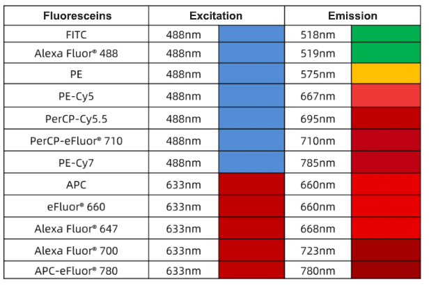
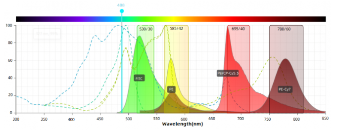
Fluoresceins can be divided into 4 types following excitation wavelength: 375 nm, 405 nm, 488 nm and 633 nm. Common excitation wavelengths are 488 nm and 633 nm fluoresceins, e.g. FITC, PE, APC, PI etc.

Fluorochrome Selection: Suitable fluorochromes depend on channel configuration and fluorochrome features(e.g. excitation spectrum, emission spectrum, brightness etc). Usually, each fluorochrome has the specific excitation and emission wavelength range. Make sure these wavelength ranges match with the channel configuration.

multicolor flow cytometry protocol

Fluorescein brightness matching: High brightness fluoresceins are recommended to antigens with low expression level. Highly expressed antigens use low brightness fluoresceins.

Minimal spectral overlap among fluoresceins: choose fluorescein combinations with less spectral overlap, e.g. FITC/PE-Cy7; choose different laser-excited fluorescein combinations: e.g. FITC/APC, PE/APC.

4. Standardized Operation and Quality Control

Standardized Operation: During multicolor antibody staining, following standardized operations can decrease experimental error and improve data reliability, including cell processing, antibody incubation and washing etc.

Quality Control: During the experiment, quality control is required to assure the accurate data. E.g. set negative and positive control to monitor the specificity and sensitivity. Meanwhile, remove interferences between different channels with fluorescence compensation.

5. Notes

Avoid to use composite fluorochromes: Although composite fluorochromes can offer multicolor choices, they are easily broken and degrade to cause unstable experiment results.

Consider autofluorescence: some cells have higher autofluorescence level and may interfere with fluorochrome detection. During choosing fluorochromes, consider the autofluorescence feature of cells and choose suitable excitation and emission wavelength range to avoid the interference of autofluorescence.

6. Conclusion

Above all, color matching of flow cytometry antibody is a complex and precise process. We need to consider antigen expression, fluorochrome features, instrument configuration and experimental operations etc. Following multicolor flow cytometry protocols and notes above can assure the accuracy and reliability of multicolor antibody staining.

7. Recommended Products

Species

Cell Populations

Flow Cytometry Antibody Combination

Cat.No

Human

T/B/NK cell populations detection

CD45-PerCP

CD3-FITC

CD16-PE

CD56-PE

CD19-APC   

PCP-30039

FITC-30004

PE-30061

PE-30008

APC-30066

Human

Th1/Th2 cell populations detection

CD3PerCP/

Cyanine5.5

CD4-FITC

IFN-γ-PE 

IL4-APC

PCP55-30004

FITC-30005

PE-30053

APC-30043

Mouse

Th1/Th2 cell populations detection

CD3-PerCP/

Cyanine5.5  

CD4-FITC

IFN-γ-PE 

IL4-APC

PCP55-30002

FITC-30001

PE-30074

APC-30026

Human

Treg cell populations detection

CD4-FITC

CD25-PE

CD127-APC   

FITC-30005

PE-30035

APC-30033

Mouse

Treg cell populations detection

CD4-FITC

CD25-PE 

FOXP3-APC

FITC-30001

PE-30017

APC-30055

Isotype Control Antibodies

Cat.No

Product Name

Host

Marker

Size

APC-30078

APC Mouse IgG2b,κ Isotype Control(MPC-11)

Mouse

APC

20T/100T

PE-30078

PE Mouse IgG2b,κ Isotype Control(MPC-11)

Mouse

PE

20T/100T

FITC-30078

FITC Mouse IgG2b,κ Isotype Control(MPC-11)

Mouse

FITC

20T/100T

APC-30077

PE Mouse IgG2a,κ Isotype Control(C1.18.4)

Mouse

APC

20T/100T

PE-30077

Mouse Anti-MPV IgG ELISA Kit

Mouse

PE

20T/100T

PCP-30076

PerCP Mouse IgG1,κ Isotype Control(MOPC-21)

Mouse

FITC

20T/100T

APC-30076

APC Mouse IgG1,κ Isotype Control(MOPC-21)

Mouse

PerCP

20T/100T

PE-30076

PE Mouse IgG1,κ Isotype Control(MOPC-21)

Mouse

APC

20T/100T

FITC-30076

FITC Mouse IgG1,κ Isotype Control(MOPC-21)

Mouse

PE

20T/100T

APC-30075

APC Rat IgG1,κ Isotype Control(HRPN)

Mouse

FITC

20T/100T

PE-30075

PE Rat IgG1,κ Isotype Control(HRPN)

Rat

APC

20T/100T

PCP-30076

Porcine Anti-MPV IgG  ELISA Kit

Rat

PE

20T/100T

FITC-30075

FITC Rat IgG1,κ Isotype Control(HRPN)

Rat

FITC

20T/100T

APC-30073

APC Rat IgG2a,κ Isotype Control(2A3)

Rat

APC

20T/100T

PE-30073

PE Rat IgG2a,κ Isotype Control(2A3)

Rat

PE

20T/100T

FITC-30073

FITC Rat IgG2a,κ Isotype Control(2A3)

Rat

FITC

20T/100T

REFERENCES

[1]Functional analysis of human NK cells by flow cytometry, PMID: 20033652.
[2]Flow cytometric analysis of T lymphocyte proliferation in vivo by EdU incorporation, PMID: 27816727.

Abstract: Ivonescimab(also called AK112 or SMT112) is the initiated humanized bispecific antibody against PD-1 and VEGF-A. It can bind with VEGF-A and PD-1 at the same time, competitively preventing their interaction with ligand. AK112 achieves one antibody with double targets and improves antitumor activity to provide a new treatment plan for patients. In clinical trials, treatment-related adverse events(TRAE) or immune-related adverse events(irAE) caused by AK112 is not observed. Overall, AK112 is found to be well-tolerated for the treatment of NSCLC. The TRAE incidence is lower than combined chemotherapy, which provides a potential treatment plan without chemotherapy in the future.

Keywords: Bispecific Antibody, Cancer Immunotherapy, Antibody Drug

1. VEGF-A Protein

VEGF-A is found in various variants. VEGFA165 is the most common one. These variants play different roles by binding with different VEGF receptors(e.g. VEGFR1, VEGFR2, VEGFR3 etc.). Receptor tyrosine kinase(RTK) is the classical VEGF receptor. VEGFR1 and VEGFR2 are mainly expressed in vascular endothelial cell(ECs). VEGFR3 is mainly distributed in lymphatic ECs to regulate generation of lymphatic vessels. VEGFA can bind with VEGFR1 or VEGFR2 according to cell type and specific functions. Compared with VEGFR2, VEGFR1 has a higher binding affinity for VEGFA. But the activity of tyrosine phosphorylation is weaker. VEGFR2 plays an important role in VEGF mediated angiogenesis and VEGF signal transduction in ECs.

bispecific antibodies - VEGF-A

2. Difference between PD-1 and PD-L1

PD-1 protein(also called CD279) consists of 288 amino acids, and exists on the surface B cell, T cell and natural killer cell. It’s up-regulated by various inflammatory stimulations in dendritic cell(DCs). PD-L1 is usually expressed by macrophage, some activated T cell and B cell, DCs, and some epithelial cells. Antigen-specific T cell plays an important role in eliminating tumor cells. Once tumor cells release tumor antigens, cancer immunity cycle will be triggered. Antigen-specific T cell initially recognizes major histocompatibility complex(MHC) presented tumor antigens. After activation and proliferation, T cell goes to the specific site along concentration gradient of chemokine. When targeting the same antigen on MHC, T cell releases IFN-γ to improve tumor killing effect. Meanwhile, The signal of TCR up-regulates the expression of PD1 on the surface of T cell. The binding between PD1 and PDL1 negatively regulates and weakens the anti-tumor function of T cell.

bispecific antibodies - PD-1

3. Anti-VEGF Drugs

VEGF is widely expressed in human tumor and cells. Especially, the subtype VEGF165 and VEGF121 are very common, which are closely related to development, metastasis, pathologic grading and prognosis of lung cancer, thyroid cancer, breast cancer, hemangioma, central nervous system tumor etc. There are five kinds of globally approved VEGF/VEGFR macromolecular drugs, including three monoclonal antibodies: Bevacizumab, Ramucirumab, Ranibizumab; and two fusion proteins: Aflibercept, Conbercept.

4. Recommended Products

4.1. Protein

Cat.No

Product Name

MW(kDa)

Host

P4791

Recombinant Human VEGFA

15.9

E.Coli

P8898

Recombinant Human CD279

41.1

HEK293 cells

P8899

Recombinant Human CD274

35-38

HEK293 cells

4.2. Antibody

Cat.No

Product Name

MW(kDa)

Type

FNab09933

VEGF antibody

45

mAb

FNab06235

PD-1 antibody

32, 50

mAb

FNab06280

CD274 antibody

50, 70

pAb

4.3. ELISA Kit

Cat.No

Product Name

Range

Sensitivity

EH3951

Human VEGF-A ELISA Kit

31.25-2000pg/ml

18.75pg/ml

EU2594

Bevacizumab(Avastin) ELISA Kit

0.313-20ng/ml

0.188ng/ml

EH4514

Human PD1/PDL1 Inhibitor Screening Assay Kit

31.25-2000pg/ml

18.75pg/ml

EH0252

Human PD-1/PDCD1 ELISA Kit

31.25-2000pg/ml

18.75pg/ml

EH5142

Anti-Human VEGF ELISA Kit

0.312-20ng/ml

0.188ng/ml

EH5110

Human anti-Bevacizumab(Avastin) antibody ELISA Kit

0.312-20ng/ml

0.188ng/ml

REFERENCES

[1]Cadonilimab, a tetravalent PD-1/CTLA-4 bispecific antibody with trans-binding and enhanced target binding avidity, PMID: 36872527.
[2]Synergistic efficacy of simultaneous anti-TGF-β/VEGF bispecific antibody and PD-1 blockade in cancer therapy, PMID: 37573354.

Abstract: Immunoprecipitation technique(IP) can purify and enrich the target protein. In the proteomics research, the protein-protein interaction can be recognized. Immunoprecipitation is applied in the protein research on relative abundance, size, up/down-regulation, stability, PTMs and interaction etc.
Keywords:    Immunoprecipitation Assay, Immunoprecipitation Technique, Proteomics

1. High Background

  • Insoluble protein: Residual insoluble proteins are in the sample. Remove the supernatant immediately after centrifugation.

  • Insufficient washing: Optimize washing buffer, improve washing efficiency by adding detergent(0.5-1%NP-40/Triton X-100/Tween-20), and increasing saline ion concentration(250mM NaCl/1-2mM DTT/β-ME), washing time as well as frequency.

  • Non-specific binding: Non-specific protein binds with the magnetic bead. Magnetic bead is insufficiently pre-blocked with BSA. Check whether the BSA is fresh. Incubate magnetic bead and 1%BSA for 1h. Wash with PBS 3-4 times before use.

  • Low specificity: use antibodies with high specificity and affinity purify.

  • Non-specific binding caused by overused antibody: try to use less antibody.

  • Overused protein in the lysate: Too many false positive proteins appear in the washing buffer. Use less sample.

  • Non-specific binding between proteins and antibodies: incubate the magnetic bead and sample before immunoprecipitation. Remove the components which can bind with the magnetic bead. Some researchers also use isotype control in lysate treatment.

  • Degradation of target protein in the sample: Perform the sample lysate at low temperature. Add excessive protease inhibitor.

  • Contamination: The high-intensity elution buffer(e.g. SDS-PAGE loading buffer) can result in the elution of various non-specific proteins and antigen. Gentle buffer(e.g. 0.1M glycine, pH2.5) can avoid the contamination.

2. Elution of Excessive Antibodies

Higher volume of antibodies, try to decrease the volume.

3. Undetected Elution of Target Protein

  • Protein expression level: The target protein in the sample is unexpressed or expressed in a low level. For low expression, use more lysate. But the non-specific binding may increase. Thus, remove the lysate before immunoprecipitation.

  • Insufficient antibody: The antibody volume is insufficient. Check and increase the volume.

  • Uneluted protein: The target protein is not eluted from the magnetic bead. Check whether the elution buffer is suitable.

  • The antibody doesn’t bind with the magnetic bead: Check whether the magnetic bead matches with antibody subtype. Store the magnetic bead properly. Avoid deterioration or drying.

  • Higher alkalinity in the lysate: Use lower alkalinity lysate. Usually, NP-40, RIPA, western or IP cellular lysate can be used.

4. Effect of Sampling Loading Order on the Recovery of Target Protein

Mixed incubation for magnetic bead, antibody and antigen sample is faster. But the purity and recovery of target protein are lower.
Indirect method: antibody binds with antigen sample first. Then add magnetic bead for incubation. The recovery of target protein is the highest; Direct method: magnetic bead binds with antibody first. Then, add antigen for incubation; For highly expressed protein, differences between the two methods are less. For lowly expressed protein, indirect method can get higher recovery of target protein.

immunoprecipitation

5. Antigen Elution Method

Denaturing elution method: This method is very efficient for WB analysis. But the eluted protein lacks of biological activity.
Acid elution method: Suitable for functional analysis of eluted protein. 0.1M-0.15 glycine (pH2.5-3) is the most efficient non-denaturing elution buffer.
Competitive elution method: For tagged target proteins, use high concentration of tag or ligand for competitive elution. The eluted protein has biological activity and antigenic fragments are not contaminated.

Antigen Elution

6. Positive and Negative Control Setting

Positive Control: Also known as input. Directly use cellular lysate or extracted protein for WB to determine whether the target protein exists in the sample.
Negative Control: Use isotype control as negative control to eliminate non-specific binding.

7. Secondary Antibody Selection

IP Capture Antibody

WB for Primary Antibody

WB for Secondary Antibody

Notes

Mouse

Mouse pAb

HRP-protein A or HRP-anti mouse IgG

If the primary antibody belongs to mouse lgG1/lgG3 subtype, the affinity of HRP-conjugated Protein A is very low. Properly decrease the dilution ratio(try to use HRP-anti mouse IgG as well); If the primary antibody belongs to mouse lgM/lgA subtype, avoid to choose HRP-Protein A due to the unconjugated feature.

Rabbit pAb

HRP-anti rabbit IgG


Rabbit

Mouse pAb

HRP-anti mouse IgG


Rabbit pAb

HRP-protein A or HRP-anti rabbit IgG light chain specific antibody

HRP-Protein A can effectively decrease heavy chain signal and the effect of light chain signal. The background is clean. Suitable for detecting most target proteins except 45-55kDa. For target proteins between 45-55kDa, HRP-anti rabbit IgG light chain specific antibody is recommended.

REFERENCES

[1]Visualization of Protein-protein Interaction in Nuclear and Cytoplasmic Fractions by Co-immunoprecipitation and In Situ Proximity Ligation Assay, PMID: 28117799.
[2]Immunoprecipitation and Western blot-based detection of protein O-GlcNAcylation in cells, PMID: 35106498.

Flow Cytometry Protocol – Part 1: Cell Surface Staining

Step 1: Cell Counting
Collect the cultured cell into 15mL centrifuge tube and count. Centrifuge at 500g for 4min and remove supernatant.

Step 2: Single Cell Suspension Preparation
Resuspend the collected cell with 1mL 0.5%BSA/PBS solution. Centrifuge at 400g for 3min and remove supernatant; Repeat twice. Resuspend the cell with 1mL 0.5%BSA/PBS solution to achieve a concentration of 1×10⁶ cell/mL. Add 100μl cell suspension into each well.

Step 3: Blocking Fc Receptors(Optional)
Incubate at room temperature for 10-20min.

Step 4: Primary Antibody Incubation
Add 100μl diluted primary antibody solution into each well, incubate at 2-8℃ for 30min.

Step 5: Washing
Centrifuge at 400g for 5min and remove supernatant. Add 200μl 0.5%BSA/PBS solution and resuspend. Centrifuge to remove supernatant. Repeat twice.

Step 6: Secondary Antibody Incubation
For non-fluorescent labeled antibodies, add 100μl fluorescent secondary antibody and resuspend. incubate at 2-8℃ for 30min in the dark area. For fluorescent labeled antibodies, jump to Step 8.

Step 7: Washing
Repeat Step 5.

Step 8: Cell Resuspension
Resuspend cell with 200μl 0.5%BSA/PBS solution. Store at 4℃ for further analysis.

Flow Cytometry Protocol – Part 2: Intracellular Staining

Step 1: Cell Counting
Collect the cultured cell into 15mL centrifuge tube and count. Centrifuge at 500g for 4min and remove supernatant.

Step 2: Single Cell Suspension Preparation
Resuspend the collected cell with 1mL 0.5%BSA/PBS solution. Centrifuge at 400g for 3min and remove supernatant; Repeat twice. Resuspend the cell with 1mL 0.5%BSA/PBS solution to achieve a concentration of 1×10⁶ cell/mL. Add 100μl cell suspension into each well. Centrifuge at 400g for 5min and remove supernatant.

Step 3: Fixation
Add 100μl cell fixation buffer into each well to resuspend cell. Incubate at 2-8℃ for 20min.

Step 4: Washing
Centrifuge at 400g for 5min. Remove supernatant and add 200μl 0.5%BSA/PBS solution to resuspend. Centrifuge to remove supernatant. Repeat twice.

Step 5: Washing
Add 100μl cell permeabilization buffer into each well to resuspend cell. Incubate at room temperature for 20min.

Step 6: Washing
Repeat Step 4.

Step 7: Blocking Fc Receptors(Optional)
Incubate at room temperature for 10-20min.

Step 8: Primary Antibody Incubation
Add 100μl diluted primary antibody solution into each well, incubate at 2-8℃ for 30min.

Step 9: Washing
Repeat Step 4.

Step 10: Secondary Antibody Incubation
For non-fluorescent labeled antibodies, add 100μl fluorescent secondary antibody and resuspend. incubate at 2-8℃ for 30min in the dark area. For fluorescent labeled antibodies, jump to Step 11.

Step 11: Washing
Repeat Step 4.

Step 12: Experimental Analysis
Resuspend cell with 200μl 0.5%BSA/PBS solution. Store at 4℃ for further analysis.

flow cytometry

REFERENCES

[1]Relevance of Antibody Validation for Flow Cytometry, PMID: 31577065.
[2]Flow cytometry-based assessment of direct-targeting anti-cancer antibody immune effector functions, PMID: 32000909.

IHC troubleshooting for endogenous peroxidase, antigen retrieval, serum blocking, antibody incubation, and DAB staining is specified below.

1. Inactivation Time and Concentration of Endogenous Peroxidase

Usually, 3% hydrogen peroxide takes shorter inactivation time and can be kept for 10min in the dark. 0.3% hydrogen peroxide can properly extend blocking time(10-30 min).

It’s suggested to prepare hydrogen peroxide in methanol which is better than double distilled water or PBS. Methanol can protect antigen and fix tissues. Overlong incubation for hydrogen peroxide can easily result in dropping.

Store the prepared hydrogen peroxide at 4ºC in the dark.

2. Antigen Retrieval Condition

Antigen retrieval is necessary for paraffin-embedded slices. Most formaldehyde fixed tissues are required for antigen retrieval before staining. Methylene bridge produced during fixing process results in protein crosslinking and shields antigen sites.

Common methods include heat induced antigen retrieval (HIER) and protease-induced epitope retrieval(PIER). Two common buffers are recommended: 10mM citrate buffer(pH 6.0) and 0.5mM Tris-EDTA buffer(pH 9.0). Suitable buffers can follow manufacturer’s instructions. Usually, Tris-EDTA buffer is applicable to most phosphotyrosine-specific antibodies. Citrate buffer applies to other antibodies.

Microwave antigen retrieval is frequently used in the lab.

3. Serum Blocking Protocol

Some spare areas in the slice can nonspecifically absorb antibodies, leading to the false-positive result. Blocking is required for the incubation of primary antibody.

Blocking with a serum homologous to the species of the secondary antibody is required. Animal’s autoantibody in the serum can bind to sites with cross reaction in tissues in advance. Otherwise, the binding with secondary antibody will result in background.

Calf serum, BSA or goat serum can be also used but should be different from the host of primary antibody.

4. Comparison of Antibody Incubation Conditions

Incubation temperature: 4ºC, room temperature, 37ºC. The best effect is 4ºC overnight.

Incubation time: related to temperature and antibody concentration. Incubate at 37ºC for 1-2 h or 4ºC overnight.

5. IHC DAB Troubleshooting 

DAB staining time is irregular and controlled under the microscope. If a light brown staining appears, washing is allowed.

DAB staining time is very short(e.g. seconds or tens of seconds). The dark chocolate-brown appears, showing the higher antibody concentration or overlong incubation. Thus, antibody concentration or incubation time should be decreased.

If the dark background appears in a very short time, the previously blocked non-specific protein may be incomplete. Then, the blocking time should be extended.

Positive staining appears after a long DAB staining time(e.g. more than ten minutes), showing lower antibody concentration, shorter incubation(4ºC overnight is better), or overlong blocking.

6. Recommended IHC Antibodies

REFERENCES

[1]Immunohistochemistry for Pathologists: Protocols, Pitfalls, and Tips, PMID: 27809448.
[2]Suggested guidelines for immunohistochemical techniques in veterinary diagnostic laboratories, PMID: 18599844.

Abstract: During western blot, some protein targets can be easily detected. However, western blot analysis always fails for other detection targets. Western blot result depends on protein molecular weight. How to conduct western blot for low molecular weight protein is specified.
Keywords:   Western Blot Protein, Western Blot Molecular Weight, Low Molecular Weight Protein

1. Western Blot Protein Loading Amount

Usually, the loading amount for each well is 30-50μg. The content of 80% protein in cells is very low. If the molecular weight is very low, 50-100μg is recommended.

2. Western Blot Gel Electrophoresis

Choose suitable gel concentration. 5% stacking gel is recommended for low molecular weight protein. Separating gel concentration is specified in the following table.

Molecular Weight(kDa)

Separating Gel Concentration

X≤10

15%

10<X≤15

13.5%

15<X≤25

12%

Low molecular weight protein running gels at higher voltage will become serrated. It’s suggested to run the stacking gel at 80V for 30 min. Later, run at 100V till the bromophenol blue (BPB) band was 1cm from the bottom of the gel.

3. Trarsmembrane

3.1. Trarsmembrane Selection

Common solid phase materials in western blot are NC membrane, DBM, DDT, nylon membrane and PVDF membrane. PVDF membrane (polyvinylidene fluoride) is recommended. Because proteins bind with PVDF membrane through hydrophobic interaction, it’s easier for antibodies to bind with hydrophilic site exposed to the liquid phase.

Two PVDF membrane for proteins with different molecular weight: 0.45μm for protein(MW>20 kDa), 0.2μm for protein(MW<20 kDa). 0.2μm can prevent proteins from directly passing through the membrane during transfer. Soak the PVDF membrane in methanol for 5min to activate the positive group and facilitate binding with negatively charged proteins. Then, put the PVDF membrane in transfer buffer.

3.2. Transmembrane Method

Conditions of wet membrane transfer are specified in the following table.

Molecular Weight(kDa)

Transmembrane Conditions

X≤10

200mA constant current for 30mins

10<X≤15

200mA constant current for 40mins

15<X≤20

200mA constant current for 50mins

The equilibration time before the transfer should be within several minutes(or even 1 min). SDS is not required for equilibrium liquid and components of transfer buffer.

4. Western Blot Ponceau Staining for Determining Protein Abundance

The stained PVDF membrane can detect western blot band size during electrophoresis. The accuracy of loading amount can be primarily evaluated by comparing the sample band in each lane.

5. Primary Antibody Dilution Ratio

Dilution ratio for low molecular weight proteins is recommended in the manual. Low dilution ratio helps antibody binding.

6. Western Blot Exposure Time

Adjust the exposure time appropriately according to signal intensity. If the band is visible in the dark place, the exposure should be within 30s-2min. If the signal is weak, the band is not clear after adding the developer. Take out of the membrane and react for a while. Then put it into the machine and add the developer again. Alternatively, a clear band can be got by extending the developing time.

Recommended Antibodies

REFERENCES

[1] Optimization of western blotting for the detection of proteins of different molecular weight, PMID: 32283940.
[2] Western blotting of high and low molecular weight proteins using heat, PMID: 26044007.

Abstract: Immunohistochemistry is the basic principle of applied immunology. Antigen-antibody reaction refers to the principle for specific binding between antigens and antibodies. During the process, the chemical reaction makes chromogenic agent (fluorescein, enzyme, metal ion, and isotope) for labelled antibodies highlighted to demonstrate the antigen in tissue cells. It’s the qualitative and relative quantitative research for antigen localization.
Keywords:       Immunohistochemistry Background Staining, IHC Background Staining, Antigen-antibody Reaction

1. Introduction

As an analytical approach, non-specific background staining usually exists. Target points in the figure below are membrane localizations. The background of the left figure is seriously stained. The staining effect of the right figure is better.

Immunohistochemistry Background Staining Effect

2. 8 Factors Influencing Immunohistochemistry Background Staining

FineTest has thousands of self-developed antibodies and rich experience in antibody test. Through thousands of slice detections, main factors for non-specific staining are found and summarized.

2.1. Antibody Quality

The low specificity of the antibody target design can result in non-specific staining. Generally, comparing with polyclonal antibodies, the specificity of monoclonal antibodies is higher. Since monoclonal antibodies can identify multiple epitopes on any antigen, monoclonal antibodies can amplify the signal of proteins with low expression level. With higher compatibility for changes in microantigen(organism polymorphism, glycosylation heterogeneity or slight degeneration ), monoclonal antibodies can be applied in detection experiment for target proteins of non-immunogenic species. During the experiment, the selection for suitable antibodies depends on real requirements. Besides, anticorrosion and prevention for repeated freezing should be noted during the storage of antibodies.

2.2. Endogenous Enzymes and Biotins

When liver, kidney or spleen tissues are used as samples, higher level of peroxidases and biotins in those tissues can cause non-specific staining during the experiment for chromogenic system using HRP or biotin-avidin. 0.9% H2O2 can be used to make endogenous peroxidase inactivated. For endogenous biotins, slices should be immersed in 25 μg/mL avidin solution for 15 min before staining. After 15min for PBS cleaning, slices can be stained. 24 mg/mL avidin can be applied to seal slices for 15min as well.

2.3. Higher Antibody Concentration

In order to obtain better chromogenic effect, the used concentration of primary antibodies should be optimized. Instructions from the supplier can be followed. The pretest is necessary to find the best working concentration.

2.4. DAB Staining Time and Deteriorated DAB

Too long DAB action time can cause background. DAB staining time always changes. During the experiment, timely microscopic examinations should be conducted. When light brown appears, DAB wash should be performed. The too short time of brown shows the antibody concentration is too high. On the contrary, the effect concentration of the antibody is too low. DAB should be stored in dry and dark place. H2O2 is just added before the usage. Immunohistochemistry background staining effects in the same parameter are showed in figures below (left: normal; right: abnormal).

Immunohistochemistry Background Staining Comparison

2.5. Dry Tissues

During the experiment process, the surface and surroundings of the sample should be kept wet. The reagent loss for causing background should be avoided. Multiple slices shouldn’t be stained at the same time and can be stained in different lots.

2.6. Immersion Time

Slices shouldn’t be immersed in buffers or retrieval buffers overnight. The experimental process should be consistent. Influences from unnecessary procedures and unknown factors should be reduced.

2.7. Insufficient Washing

Incubated antibodies should be fully washed. Before using PBS, pH value should be defined. Tween-20 can be added to increase washing strength if necessary.

2.8. Sealing

Serum diluents are used to seal for immunohistochemistry. To remove the non-specific adsorption caused by secondary antibodies, non-immunized animals’ serums can be used. E.g. If the secondary antibody is goat anti rabbit, non-immunized goat serum should be chosen. It’s suggested to use PBS to dilute 3-10% solution where slices are incubated for 10-30min under 37°C. It’s not necessary to clean after sealing. Moreover, after the tissue is fixed, part free aldehyde group may be residual. The non-specific binding may happen during incubating antibodies. Then, 0.3M glycine can be used to seal.

3. FineTest Antibodies

FineTest offers high-quality validated antibodies, including polyclonal antibodies, monoclonal antibodies, labelled secondary antibodies, internal control/tag antibodies, acetylated/phosphorylated antibodies etc. These antibodies are mainly applied in intracellular and extracellular protein identification, quantitative/positioning research, western blot, immunofluorescence, immunohistochemistry, FACS, immunoprecipitation/immunocoprecipitation. FineTest antibodies are strictly tested to ensure the purity and quality. FineTest is always committed to contributing to advance your scientific research.

REFERENCES

An improved protocol of biotinylated tyramine-based immunohistochemistry minimizing nonspecific background staining, PMID: 12502763.

Abstract: Before the invention of heat induced antigen retrieval solution (HIER), protease-induced epitope retrieval(PIER) is the most commonly used method. Many enzymatic techniques are ever used, including trypsin, pronase, ficin, pepsin and proteinase k antigen retrieval etc.
Keywords:  Antigen Retrieval Solution, Antigen Retrieval, Enzymatic Antigen Retrieval

1. Protease-induced Epitope Retrieval

The mechanism of PIER is probably to stop the protein crosslink produced during formalin fixed process by enzymatic digestion. However, the stopping is non-specific and has negative influence on some antigens. The effect of PIER depends on the enzyme concentration and type, incubation parameters(time, temperature, pH) and fixation time. The enzymatic digestion time and fixation time are negatively correlated.

PIER Antigen Retrieval Solution

2. PIER Antigen Retrieval Solution & Optimization

FineTest and other laboratories usually optimize induced epitope retrieval condition of a few enzymes instead of trying many enzymes. FineTest adopts the ready-to-use protease K solution which has good activity at room temperature and can be applied in automated immunostaining device.

Disadvantages of PIER:

  • Not suitable for most antigens;

  • May result in morphological changes;

  • May destroy antigenic peptide

3. Enzymatic Antigen Retrieval Solution

Three enzymatic digestion methods are specified below:

3.1. Trypsin

  • Immerse the slide into 0.1% trypsin solution (pH7.8), incubate for 10-30 min at 37℃;

  • Rinse completely by running water;

  • Immunohistochemical staining

3.2. Protease

  • Drop 0.05%-0.5% (w/V) warm protease XXIV on the tissue slice and incubate for 10-25min at 37℃;

  • Rinse completely by running water;

  • Immunohistochemical staining

3.3. Pepsin

  • Immerse the slide into 0.4 % (w/V) pepsin solution;

  • Incubate for 10-25min at 37℃;

  • Rinse completely by running water;

  • Immunohistochemical staining

4. FineTest Antibody

Browse a list of FineTest antibody.

REFERENCES

Effect of Antigen Retrieval Methods on Nonspecific Binding of Antibody-Metal Nanoparticle Conjugates on Formalin-Fixed Paraffin-Embedded Tissue.

Abstract: The cross-linking during the tissue fixation changes tertiary and quaternary structures of many antigens. Specific antibodies can hardly detect. Antigen retrieval methods aim to restore the lost antigenicity and theoretically restore protein conformation before the fixation. About 85% formalin-fixed antigens should be optimized for immune reaction by the antigen retrieval method.
Keywords: Antigen Retrieval, Antigen Retrieval in IHC, Antigen Retrieval Methods

1. Antigen Retrieval Protocol

The necessity of antigen retrieval depends on antigens ready for detection and used antibodies. Compared with monoclonal antibodies, polyclonal antibodies have high sensitivity and can more easily detect antigens without antigen retrieval. Currently, there are two kinds of antigen retrieval methods: heat induced antigen retrieval (HIER) and protease induced epitope retrieval (PIER).

2. Detergents and Chaotropic Substances

Detergents and chaotropic substances are usually together used with HIER or PIER. The efficiency is lower than HIER and PIER.

2.1. Antigen Retrieval Methods Using Detergents

Detergents can form particles in the aqueous solution and decrease surface tension of water. Hence, they are also surfactants. Detergents simulate lipid bilayer environment and can dissolve membrane protein. Then, detergents micelle consisting of lipid, detergents and proteins form mixed particles (Usually, a particle contains a protein molecule). Common detergents in IHC are non-ionic. These substances are suitable for breaking lipid–lipid interactions and lipid–protein interactions without damaging protein–protein interactions. Thus, detergents are not denaturants. Non-ionic detergents mainly include TritonR-X100, Tween20, saponin, BRIJR and NP-40. Generally, they’re added into wash buffer(e.g.  0.05% V/V Tween 20 ).

Antigen Retrieval Methods

2.2. Antigen Retrieval Methods Using Detergents

Dispersants cover guanidine hydrochloride, sodium sulfocyanate and cesium. They’re considered as protein denaturants. The partial opening or reversal of formalin results in protein crosslinking by opening protein complex. Usually, the efficiency of using detergents or dispersants is low.

3. FineTest Antibody

Browse a list of FineTest antibody.

REFERENCES

[1] How does antigen retrieval work?
[2] Antigen retrieval techniques: current perspectives

Abstract: Activity of some endogenous enzyme and other endogenous substances plays an important role in immunohistochemistry. Then, what are causes of background staining or non-specific binding of primary antibody?
Keywords: Immunohistochemical Staining, Non-specific Binding of Antibodies, Immunologic Test

1. Antibody Titration

Higher titer of primary antibody is one of the reasons for background staining. Reasonable antibody titer can produce strong specific signal with low or none background staining. Each kind of antibody should carry out standard titer by chessboard titration. At least, several titer tests are required. Many variables can influence the best primary antibody titer, including incubation time and temperature, antibody retrieval parameters (method, time, temperature, pH, chemical components), buffer (PBS or TBS, pH) and detection method (sensitivity, specificity, time, temperature).

IHC Background Staining for Liver Cancer

Figure 1. IHC Test for Liver Cancer.

2. Specific Cross Reaction of Antigen

This term defines the specific binding between antibodies with other antigens existing in histiocyte in addition to target antigen. Different cell or tissue has the same antigenic epitope of the antibody. E.g. CD79a monoclonal antibody can not only bind with CD79a molecule on B lymphocyte, but also strongly label smooth muscle cell. CD15 is applied for staining Hodgkin’s lymphoma Reed-Sternberg cells. However, there is cross reaction between CD15 antibody and macrophages. A special specific cross reaction is called cross-species reaction. The same antigen (epitope) existing in the cytokeratin or vimentin of multiple species(e.g. dog, cat, horse, anti porcine coronavirus monoclonal antibody can react with ferret/dog/cat/pig coronavirus.) Secondary antibodies in immunohistochemistry may also react with tissue antigen (E.g. Rabbit anti rat second antibody may react with mouse tissue antigen). Using immunoglobulin for the same species of the tissue to be tested adsorbs the second antibody in order to remove the reaction. (E.g. if the rat secondary antibody is used in immunohistochemical staining of mouse tissue, mouse immunoglobulin or serum protein is required for removing the cross-species reaction.)

IHC Background Staining for Kidney Cancer

Figure 2. IHC Test for Kidney Cancer(20X).

3. Non-specific Cross Reaction of Antibody

The primary or secondary antibody binds with the irrelevant protein by non immune mechanism. Background can occur by tissue fixation or antigen retrieval resulting in conformational changes of protein.

IHC Background Staining for Colon Cancer

Figure 3. IHC Test for Colon Cancer.

Browse a list of FineTest IHC antibodies.

REFERENCES

Non-specific binding of antibodies in immunohistochemistry: fallacies and facts.

Abstract: In immunohistochemistry test, the most common questions are influences of background and how to explain experimental result. There are many reasons for background staining. Two main reasons are specified in this article: endogenous enzyme activity and activity of other endogenous substances.
Keywords: Immunohistochemistry Test, Immunostaining, Enzymology

1. Endogenous Enzyme Activity

1.1. Endogenous Peroxidase Activity

Endogenous peroxidase activity naturally exists in in erythrocytes(pseudoperoxidase), granulocytes(myeloperoxidase) and neurons. It can react with DAB to produce brown products and are hard to be distinguished from specific immunostaining. Pretreatment with H2O2 dilution(0.03%-3%, including 0.1% sodium azide) can reduce or remove pseudoperoxidase activity of frozen sections of erythrocytes and peroxidase activity of myeloid cells. Regular paraffin-embedded tissue slices and hemin slices with massive haemorrhage or acidity require higher concentration H2O2(3% H2O2 solution and 0.1% sodium azide). Alternatively, the endogenous enzyme activity can be removed by extending incubation time in lower concentration solution. H2O2-methanol is not recommended for regular use, especially for testing cell surface antigens.

Methanol has a bad influence on antigenicity and immunoreactivity. We should avoid to use it. However, when using H2O2-methanol, the incubation time should be within 15min.

1.2. Endogenous Alkaline Phosphatase(AP)

Alkaline phosphatase in mammalian tissues has intestinal or non-intestinal isozyme. The background staining will happen in the alkaline phosphatase method. 1mmol/L levamisole can inhibit non-intestinal alkaline phosphatase isozyme. However, isoenzyms of bovine intestinal alkaline phosphatase is unaffected as a reporter in alkaline phosphatase immunostaining method. 1% acetic acid can block the isozyme but destroy some antigens at the same time.

2. Activity of Other Endogenous Substances

2.1. Endogenous Avidin/Biotin Activity

Endogenous biotin widely exists in mammalian tissues and especially is rich in tissues like liver, lung, spleen, adipose tissue, kidney, brain etc. After the fixation of the tissue by formalin, background caused by endogenous biotin greatly decreases but increases in frozen sections. Strong ionic strength of alkaline avidin has high adsorption capacity for cellular molecules with opposite charges and non specific binding occurs, E.g.: glycosaminoglycans(GAGs) in nucleic acids, phospholipids, mast cell cytoplasm. The isoelectric point of avidin is 10. Avidin is alkaline in immunostaining buffer with pH close to neutrality and carries a positive charge. The strong heat-induced antigen retrieval will get endogenous biotin in the formalin-fixed tissue exposed. However, avidin in the detection system can bind with it and present strong background required for inhibition. Preparing avidin-biotin complex (ABC) by pH9.4 biotinylated solution can prevent the non-immune binding.

2.2. Endogenous Immunoglobulin Activity

In immunohistochemistry, the binding between the primary antibody and immunoglobulin on the tissue surface can cause background. Before the incubation of the primary antibody, the non-specific blocking reagent should be used, E.g.: 5% bovine serum albumin(BSA) can block the binding between the primary antibody and non-specific antigen in the tissue.

3. IHC Test Results by FineTest Products

Immunohistochemistry Test for Lung Cancer

Figure 1.  lung cancer 20x cyt+++.

Immunohistochemistry Test for Cervical Carcinoma

Figure 2.  cervical carcinoma 20x.

Browse a list of FineTest IHC Products.

REFERENCES

[1] Background staining of visualization systems in immunohistochemistry: comparison of the Avidin-Biotin Complex system and the EnVision+ system.

[2] Isozymes of bovine intestinal alkaline phosphatase.

[3] Inhibition of endogenous tissue alkaline phosphatase with the use of alkaline phosphatase conjugates in immunohistochemistry.

Abstract: Currently, many kinds of enzymes are applied in immunostaining. Horseradish peroxidase(HRP) and alkaline phosphatase(AP) are the two enzymes most widely used in enzyme labeling methods.
Keywords:  Immunohistochemistry, Enzymology, Horseradish Peroxidase(HRP), Alkaline Phosphatase(AP), Immunostaining

1. Horseradish Peroxidase(HRP)

The substrate of HRP is hydrogen peroxide. Peroxidation products oxidize chromogen and precipitate in the binding site of antibodies. 3,3′-Diaminobenzidine tetrahydrochloride (DAB) is the most commonly used chromogens,  which can produce brown precipitation insoluble in organic solvents. If the activity of endoperoxidase or melanin content is higher, DAB isn’t the best choice. Then, other chromogens or alkaline phosphatase should be considered. Another chromogen of peroxidase 3-Amino-9-ethylcarbazole (AEC) produces red precipitation soluble in organic solvents and should be sealed in water-soluble liquid. Other chromogens can also produce similar red precipitation insoluble in organic solvents. 4-Chloro-1-naphthol can produce blue precipitation soluble in organic solvents. Currently, several different experimental methods can enhance the colour of DAB end products.

2. Alkaline Phosphatase(AP)

Alkaline phosphatase is separated from the intestinal tissue of calf. Basic principles are as follows. By hydrolyzing phosphorous salts containing naphthol substitutes, insoluble naphthol derivatives are produced. Then, the derivatives bind with suitable diazonium salts and produce colored insoluble azo dyes in the active site of the enzyme. The most frequently used chromogens are BCIP/NBT(blue, stable medium), Fast Red (red, water soluble sealing reagent) and New Fuchsin (magenta, water soluble sealing reagent). Alkaline phosphatase is suitable for immunocytochemistry test for cell samples or tissue samples with higher endoperoxidase. Alkaline phosphatase and horseradish peroxidase can be jointly applied in double immunohistochemical staining.

3. FineTest IHC Products

FineTest has rich experience in HRP enzyme labeling. Applications of HRP conjugated secondary antibodies in IHC staining are shown in the figures below.

gastric cancer immunostaining

Figure 1. Gastric cancer 20×.

kidney immunostaining

Figure 2.  Kidney 10×.

colon cancer immunostaining

Figure 3. Colon cancer.

Table 1. Selected FineTest IHC Products

Cat No.

Product Name

IHC0001

Fine Test SABC KIT

IHC0002

Fine Test SABC KIT

IHC0003

Fine Test SABC KIT

IHC0004

Fine Test SABC KIT

IHC0005

DAB Substrate Kit

IHC0007

Rabbit-DAB (Poly-HRP) Detection IHC Kit

IHC0008

Mouse-DAB (Poly-HRP) Detection IHC Kit

REFERENCES

[1] Horseradish peroxidase: a modern view of a classic enzyme.
[2] Alkaline Phosphatase: Discovery and Naming of Our Favorite Enzyme.
[3] Multiple Antigen Immunostaining Procedures.

Abstract: Tissue preparation of immunohistochemical reaction has been mentioned in the previous article about immunohistochemistry, which greatly determines whether the experiment can be successful. Let’s learn details and methods of five kinds of tissue preparation.
Keywords: Tissue Preparation for Immunohistochemistry, IHC

1. Slides in Tissue Preparation

High quality immunohistochemical result requires the reasonable combination of tissue slices and slides in order to prevent reagent aggregations and pseudostaining caused by tissue adhesion. Commercial slides specially used to perform immunohistochemical staining are standardizedly coated. Please find two coating protocols for regular slides used in immunohistochemical staining.

1.1. Poly-L-Lysine Coating Slides Protocol

  • Prepare 0.1%(w/V) poly-l-lysine solution(Sigma P-8920);

  • Immerse the slide in the solution;

  • Dry at room temperature;

  • Store at room temperature.

1.2. APTS Coating Slides Protocol

  • Prepare acetone solution containing 2%(V/V) silane(Sigma A-3648);

  • Immerse the slide in the acetone for 1-5min;

  • Immerse the slide in the acetone solution containing 2% silane for 1-5min;

  • Continuously wash the slide in the acetone solution twice for 5min;

  • Dry and store at room temperature.

2. Preparation of Cell Smear/Centrifuged Cell Smear

  • Prepare cell scraping smear/centrifuged cell smear with slide/poly-l-lysine coating slides;

  • Dry at room temperature for 30min;

  • Fix in pre-cooling and fresh acetone for 20min at -20℃;

  • Air-dry at room temperature for 15min at least;

  • Stain cell scraping smear/centrifuged cell smear.

3. Frozen Sections in Tissue Preparation

  • Cut four 7μm unfixed frozen sections;

  • Put sections on slide/poly-l-lysine coating slides;

  • Air-dry sections in the air for 30min;

  • Put sections in fresh acetone pre-cooled to 4℃ and fix for 20min;

  • Air-dry sections for at least 15min at room temperature;

  • Stain sections.

4. Notes for Cell Smear and Frozen Sections

  • Fresh acetone can be replaced by 100% ethanol; The selection of fixative depends on the target antigen to be stained in the tissue;

  • Unstained slides can be coated in aluminum foil and stored at -20℃. When ready for staining: open and recover aluminum foil to room temperature. Fix for at least 30s with the same fixative before the storage. Air-dry slices and then rehydrate for 5min in buffer. Finally the staining is performed;

  • The stability of unstained slides depends on characterization of antigens; Some antigens are very unstable and should be stained after smearing.

5. Paraffin Sections

  • Fix the tissue in 10% formaldehyde for 12-48h(other fixatives may be also suitable.) Fixation time depends on the thickness and temperature of tissues as well as properties of antigens ready for detection;

  • Paraffin-embedded;

  • Cut 3-5μm section;

  • Put the section on slide/poly-l-lysine coating slides and perform the water bath without the adhesive;

  • Blot up the water completely along the edge of the section;

  • Put the section in 60℃ oven for 30-60min;

  • Stain the section after being taken off paraffin.

Notes

  • Extending the drying time(e.g. Overnight at room temperature)for tissues rich in fat(e.g. brain, breast) may enhance the adhesion of tissue slices;

  • Over 60℃ drying temperature may be harmful to the detection of some antigens or increase background staining;

  • In any storage condition, extending the storage time of paraffin section may decrease immunoreactivity of some antigens. The following antigens are easy to be degraded: estrogen receptor, progesterone receptor, proteins related to cell cycle regulation(e.g. Ki-67、PCNA、cyclinD1、bcl-6) and thyroid transcription factor-1. It’s unsuitable to store paraffin sections for a long time;

  • If the staining of the archival paraffin section is inconsistent, fresh sections should be used to repeat the experiment.

6. Cell Block

This step is suitable for the difficult paraffin embedding because the tissue block is too small. A reagent that forms so-called cell blocks is essential. Four different types of preparing cell blocks are below.

6.1. Agar Cell Block Preparation

  • Take 20ml precipitation sample at the bottom and perform the centrifugation at 3000r/min for 5-10min;

  • Blot up the supernatant and fix centrifugal sediments for 1h by 10% neutral formalin;

  • Blot up the liquid;

  • Add a little melted agar and shake the test tube for the complete mixture. Then perform the centrifugation at 3000r/min for 5-10min. At present, agar has been solidified;

  • Take out the agar cell block and put it into dehydration box with the suitable cutting size. Then prepare paraffin-embedded blocks as usual.

6.2. Albumen Cell Block Preparation

  • Take 20ml precipitation sample at the bottom and perform the centrifugation at 3000r/min for 5-10min;

  • Add lml egg white and then gently scrape cells at the bottom of the tube with flat bamboo stick, making cells form the cluster and suspended in egg white. Similarly, perform the centrifugation at 2000r/min for 5min;

  • Remove redundant egg white and gently add 2ml 10 % neutral formalin fixative solution along the tube wall. The egg white is immediately solidified to get cell cluster. Then, shake gently to make cell cluster suspended and fixed in the fixative solution overnight. Gently take out the cell cluster and wrap it with the embedding paper. Finally, put it into dehydration box and prepare paraffin-embedded blocks as usual.

6.3. Direct Cell Block Preparation(Applicable for A Large Amount of Cell)

  • Take 20ml precipitation sample at the bottom and perform the centrifugation at 3000r/min for 5-10min;

  • Blot up the supernatant. Then, gently add 95% alcohol along the tube wall and perform the centrifugation at 3000r/min for 5min. The standstill time should be no less than 1h;

  • Gently take out sediments with a bamboo stick and wrap it with the embedding paper;

  • Put it into dehydration box and fix it by 10% neutral formalin for 1h. Then prepare the paraffin section as usual.

6.4. Glutaraldehyde Cell Block Preparation

  • Take 20ml precipitation sample at the bottom and perform the centrifugation at 3000r/min for 5-10min;

  • Blot up the supernatant. Add 10ml 3% glutaraldehyde for the complete mixture. Then, perform the centrifugation at 3000r/min for 5-10min;

  • Gently take out sediments with a bamboo stick;

  • Put it into dehydration box and fix it by 10% neutral formalin for 1h. Then prepare the paraffin section as usual.

Abstract: Immunohistochemistry (IHC) is a technique for detecting antibodies in tissue slices using specific antibodies. Compared with other techniques, IHC has its unique feature in protein identification. The integration of antigen detection and its localization in tissues or cells is important for the research on cell function of normal or abnormal tissues. IHC integrates three important areas: morphology, histochemistry and immunology. The detection of antigens in tissue slices will be mainly introduced. The staining for cell slide is briefly introduced as well (This is called immunocytochemistry). Find how to standardize new immunohistochemical method.
Keywords:   Immunohistochemistry, IHC, Immunohistochemical Method, Antigen Test, Antigen Retrieval, FineTest Antibodies

1. Evolution History of Methodology

In the past, immunohistochemical method uses fluorescent labeling and frozen slices. With the technical advancement, immunohistochemistry can be applied in formalin-fixed and paraffin-embedded tissue slices. Then, antigen detection is related to morphology. The archived paraffin tissue block is also used.

In immunohistochemical method, the binding of antibodies and antigens in tissues is shown by immunohistochemical staining. Since the fixation by Formalin can prevent antigen detection. Currently, many developed antigen retrieval methods can solve the problem. The reaction sensitivity is very important for detecting some microantigens.

During the past ten years, many highly sensitive immunohistochemical methods were found. Different combinations of enzyme-substrate-chromogen can generate various colors. Thus, various kinds of antigens can be detected on the slice in the same time.

2. A New Practical Method for Standardized IHC

It’s difficult to standardize a new immunohistochemistry, because many factors can influence the result. Most antibodies in the market are just tested in human tissues but they are hardly detected in other species. The product manual offered by the antibody manufacturer is very important for using antibodies, which specifies whether the antibody can detect the antigen of a certain species, the antigen shown by western blot using immunohistochemical method, and the antigen in frozen slices etc. However, such information is unavailable in many cases. Then, researchers have to develop a standardized method for detecting new antibodies. The research programme on formalin-fixed/paraffin-embedded tissue block is outlined below.

Methodology:

  • Select the tissue applicable for positive control; It’s better to include known adjacent tissues of unexpressed antigen pending for detection;

  • Processes like fixation and embedding etc of control tissues should be the same with tissues ready for detection. Control tissues and tissues ready for detection come from the same species. If tissues of other species are found for reacting with the antibody, it’s recommended to set another control group;

  • Dilute primary antibodies(Usually, antibodies diluted in 1:16 fits for immunohistochemical staining.);

  • Three groups of slides are prepared for identifying the best antigen retrieval method(AR): The first group is not involved in any antigen retrieval; The second group uses the enzyme antigen retrieval(e.g. Proteinase K); The third group uses pH6.0 citric acid buffer for heat-induced antigen retrieval;

  • Perform the immunohistochemical test according to a standard process. Then select the incubation time and temperature of primary antibodies based on known antibody information. Without the information, incubate the antibody for 60min at room temperature;

  • At the end of IHC detection, observe the staining effect of the slide.

R brain 20x Nuc+++

3. Notes

  • If the specific staining is successful, it’s required to further optimize concentration, incubation time & temperature and antigen retrieval method of primary antibodies in order to obtain the best SNR;

  • If the staining fails, it’s required to further regulate concentration, incubation time & temperature (4℃ overnight or even longer incubation time) and antigen retrieval method of primary antibodies(Use other enzymes or different pH buffers);

  • The antibody may still not detect antigen finally due to the following factors: influences of tissue fixation for antigens, species differences of antigens, lack or low expression of recognized epitope of antibodies etc.

H liver 20x cyt+++

4. Tissue Preparation

In the next article, tissue preparation will be specified. FineTest offers IHC antibodies as well. Browse a list of 10,000+ FineTest Antibodies.

Abstract: In the previous article, we have preliminarily learned about immunohistochemistry. Then, antibody preparation involves the immunization of purified antigens (e.g. mouse, rabbit, goat, horse etc).
Keywords:   Immunohistochemistry, Tissue Preparation, Antibody Preparation

1. Immunohistochemistry Antibodies

Various animals can be applied to produce polyclonal antibodies, especially rabbit, horse, goat and chicken etc. Polyclonal antibodies have a high affinity and wide reactivities. However, compared with monoclonal antibodies, the specificity is lower. Because polyclonal antibodies can bind with multiple antigenic epitopess in fixed tissues, the sensitivity is improved. Usually, antibodies which can recognize strong immunogenicity and weak antigenic epitopes exist in polyclonal antibodies reagents. Hence, polyclonal antibodies may be very inconsistent. The binding of multiple epitopes is a advantage.(It can bind with several antigenic epitopes in the same protein. Antigens can be more likely to be detected.) The possibility of cross reaction with other proteins which have similar antigenic epitopes will be increased, resulting in the false positive.

Immunohistochemistry Result_1

Monoclonal antibodies are usually produced by mice. The technology is developed by Kohler and Milstein. Compared with polyclonal antibodies, the advantage of monoclonal antibodies is high specificity which decreases the cross reaction with other antigen(It’s impossible to completely remove the reaction). If the cross reaction appears, the possible reason is that antigenic epitopes targeted by monoclonal antibodies just consist of several amino acids. However, the antigenic epitope exists in various proteins and polypeptides. Sometimes, it’s very hard to confirm the non-specific immune response is caused by common antigenic epitopes(cross reaction) or protein crosslinks when the tissue is fixed by aldehyde fixatives. In some situations, the background colour of monoclonal antibodies caused by non-specific immunoglobulins can decrease ascites or disappear in the culture supernatant of hybridoma cells.

2. Tissue Preparation for Immunohistochemistry

The tissue preparation determines whether the immunohistochemical reaction can succeeds to a great extent, which mainly depends on antigen types to be tested in some ways. E.g. During the fixation of formaldehyde, some antigens will be destroyed. Hence, these tissues should be frozen or fixed by other stationary liquids. A principle for testing antigens should be followed: samples are required to be collected and treated before the decomposition of tissues.

Immunohistochemistry Result_2

3. Tissue Fixation for Immunohistochemistry

Tissues which are not instantly frozen should be fixed at once. Tissue fixation is very necessary. Reasons are as follows(functions of tissue fixation):

  • Preserve cell components like soluble protein and structural protein etc;

  • Prevent the degradation and translocation of cell components like antigen and some enzymes;

  • Stabilize cell components to reduce harmful effects of next steps;

  • Help traditional immunohistochemistry staining and immunostaining. Two types of fixatives are mainly applied in histopathology: crosslinking fixatives(non-coagulant) and coagulant fixatives.

Formaldehyde is the golden fixative in regular histochemistry and immunohistochemistry. It can preserve most polypeptide and general structure of organelle, and can also react with nucleic acid. However, carbohydrates are not influenced. When the calcium ion is contained, it’s the good protective reagent for lipid. The basic fixing mechanism of formaldehyde: formalin and active amido without charges (-NH or -NH2) form a addition product. Finally, modifications of the methylene bridge (crosslinking) are formed in the tertiary and quaternary structure of proteins. Formaldehyde fixation is a gradual process with dependent time and temperature. Over fixation will result in the immunohistochemical false negative outcome caused by over crosslinking. However, insufficient fixation will generate unexpected results. There is no the most suitable fixing time for the fixation of each antigen.

Immunohistochemistry Result_3

Many substitutes of formalin are some coagulant fixatives, which can destroy hydrogen bond to precipitate proteins instead of protein crosslinking. The most typical non-crosslinking fixative is ethanol. Other fixatives applied in immunohistochemistry are glyoxal(dialdehyde), the mixture of glyoxal and ethanol, 4% paraformaldehyde and zinc formalin etc. Compared with formalin, their effects on protein conformational changes are not obvious. It’s suggested to follow instructions recommended by the antibody manufacturer to find the most suitable fixing method. For immunohistochemical primary antibodies, if staining results for frozen section are obtained, the staining of fixing tissue is pending for testing. It’s suitable to use 4% paraformaldehyde or zinc formalin for fixing paraffin-embedded tissues during testing the staining of fixing tissue. This method is good for using paraffin-embedded tissues. Because different fixatives variably influence immune response, the tissue paraffin fixed by non formaldehyde fixative should be properly labelled before archiving.

4. FineTest IHC Antibodies

FineTest offers IHC antibodies for research use. Browse a list of FineTest antibody.