Antibodies—also known as immunoglobulins (Ig)—are glycoproteins produced by B cells in response to foreign antigens. In the immune system, they function as critical mediators of antigen neutralization and immune signaling. In biomedical science, antibodies are essential reagents used across a wide range of experimental techniques, including Western blotting (WB), immunohistochemistry (IHC), flow cytometry (FC), and enzyme-linked immunosorbent assays (ELISA).
A comprehensive understanding of antibody structure is fundamental for scientists seeking to interpret experimental results, engineer novel antibody formats (e.g., bispecifics, antibody-drug conjugates), and troubleshoot technical issues.
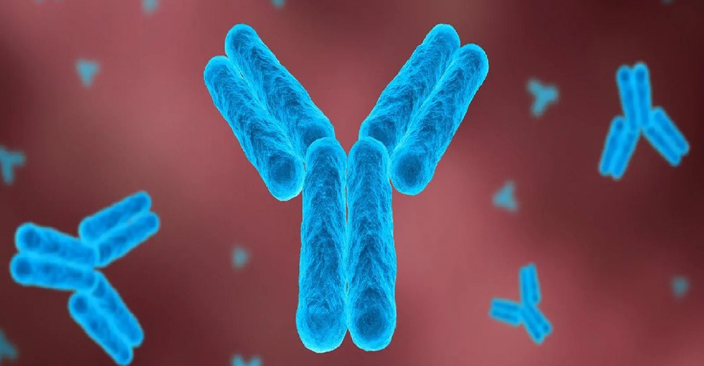
Overview of the Antibody Molecule
A typical antibody is a Y-shaped molecule consisting of two identical heavy chains (~50 kDa each) and two identical light chains (~25 kDa each). These chains are linked via disulfide bonds and stabilized by non-covalent interactions. The molecule is functionally and structurally divided into three key regions:
- Fab (Fragment antigen-binding)
- Fc (Fragment crystallizable)
- Hinge region
Each of these plays a unique role in the antibody’s interaction with both antigens and the immune system.

The Fab Region: Antigen Binding and Variable Domains
The Fab region is located at the “arms” of the Y-shaped structure and is responsible for antigen recognition. Each Fab fragment contains:
- One variable domain of the heavy chain (VH)
- One variable domain of the light chain (VL)
- One constant domain of the heavy chain (CH1)
- One constant domain of the light chain (CL)
The variable regions (VH and VL) are what make antibodies so powerful and versatile—they contain the Complementarity-Determining Regions (CDRs) that directly bind to specific epitopes on the antigen.
The Fab fragment can be isolated enzymatically (e.g., papain digestion) for therapeutic or diagnostic purposes where antigen binding without Fc effector function is desired.
The Fc Region: Effector Functions and Immune Signaling
The Fc region (the “tail” of the antibody) consists of the CH2 and CH3 domains of the two heavy chains and is essential for immune effector functions. It mediates:
- Binding to Fc receptors (FcγRs) on immune cells, enabling antibody-dependent cell-mediated cytotoxicity (ADCC) and phagocytosis
- Complement activation via the classical pathway, leading to opsonization and pathogen lysis
- Half-life extension by interaction with neonatal Fc receptors (FcRn)
Engineering of the Fc region (e.g., glycosylation modification, Fc silencing) is a major area in antibody therapeutics, especially to modulate ADCC or increase serum half-life.
The Hinge Region: Structural Flexibility
The hinge region connects the Fab arms to the Fc tail and imparts flexibility, allowing the antibody to adapt to:
- Spatially separated epitopes on multivalent antigens
- Conformational changes during antigen binding
- Cross-linking of antigens to form immune complexes
This region contains cysteine residues for inter-chain disulfide bonding and proline-rich sequences that influence structural dynamics.
The CDRs: Antigen Recognition Hotspots
Each variable domain (VH and VL) includes three CDRs:
- CDR1, CDR2, CDR3 (numbered from the N-terminus)
CDR3 is generally the most variable and is critical for determining specificity. The six CDRs (3 on the heavy chain and 3 on the light chain) together form the paratope, the actual antigen-binding site.
The flanking regions—known as framework regions (FRs)—provide structural stability and affect CDR conformation. Subtle mutations in FRs can enhance or impair binding without altering CDR sequences.
Antibody Isotypes and Subclasses
In humans, there are five major isotypes: IgG, IgA, IgM, IgE, and IgD. Each differs in Fc structure and function.
| Isotype | Structure | Key Functions |
| IgG | Monomer | Opsonization, complement activation, ADCC, long serum half-life |
| IgA | Dimer (secreted) | Mucosal immunity (saliva, tears, gut) |
| IgM | Pentamer | First responder, potent agglutination and complement activation |
| IgE | Monomer | Allergy and parasite immunity (binds FcεRI on mast cells) |
| IgD | Monomer | Primarily functions as a B-cell receptor (function still under investigation) |
IgG is further subclassed into IgG1, IgG2, IgG3, and IgG4, each with distinct effector profiles and pharmacokinetics.
Structural Variants: scFv, Fab’, F(ab’)2, and More
In modern antibody engineering, structural variants are commonly used for diagnostics, research, and therapeutics:
- scFv (Single-chain variable fragment): Compact format combining VH and VL via a flexible linker
- Fab and F(ab’)2 fragments: Lacking Fc region; used to prevent Fc-mediated interactions
- Nanobodies (VHH): Derived from camelids; extremely small and stable
- Bispecifics: Two antigen-binding sites for different targets, often engineered into IgG-like formats
- Fc-fusions: Fusion of Fc domain with a ligand, receptor, or peptide for enhanced stability
Each variant serves unique experimental or clinical needs.
Implications for Antibody-Based Assays
Understanding antibody structure allows researchers to:
- Design better experiments: Know whether Fc interactions might cause background staining in IHC or FC
- Optimize detection systems: Use Fc-blocking reagents to reduce non-specific binding
- Interpret band patterns: Multiple bands in Western blot may indicate post-translational modifications or antibody cross-reactivity due to suboptimal CDR specificity
- Select appropriate reagents: Fragment antibodies (e.g., F(ab’)₂) can be useful when Fc-mediated binding skews results
Structural Prediction and Engineering in 2025
In 2025, deep learning tools like AlphaFold2-Multimer, IgFold, and AntiFold have made it possible to accurately predict antibody structures, including CDR loop conformations. These models help:
- Design antibodies with desired specificity
- Predict binding interfaces and epitope accessibility
- Engineer humanized or Fc-silenced variants
Structure-guided design is now a standard step in the development of recombinant antibodies and antibody-drug conjugates.
Conclusion
Antibody structure is not merely academic—it directly informs function, assay performance, and therapeutic efficacy. From Fab-mediated specificity to Fc effector engagement and CDR diversity, each component of the antibody plays a role in determining outcome.
At KinesisDx, our antibodies are rigorously engineered and validated with structure-function relationships in mind. Whether you’re working on a diagnostic platform, basic research, or therapeutic exploration, understanding antibody structure is the first step in achieving reproducible and biologically meaningful results.
Works Cited (MLA Format)
Wikimedia Commons. “Antibody Structure.” Wikimedia Commons, https://commons.wikimedia.org/wiki/File:Antibody_structure.svg.
Janeway, Charles A., et al. Immunobiology: The Immune System in Health and Disease. 5th ed., Garland Science, 2001.
Alberts, Bruce, et al. Molecular Biology of the Cell. 6th ed., Garland Science, 2014.
Harlow, Ed, and David Lane. Antibodies: A Laboratory Manual. Cold Spring Harbor Laboratory Press, 1988.
Uhlén, Mathias, et al. “A Proposal for Validation of Antibodies.” Nature Methods, vol. 13, no. 10, 2016, pp. 823–827.
Lefranc, Marie-Paule, et al. “IMGT Unique Numbering for Immunoglobulin and T Cell Receptor Variable Domains.” Developmental and Comparative Immunology, vol. 27, no. 1, 2003, pp. 55–77.
Ruffolo, Justin A., et al. “Transfer Learning Enables Predictions in the Antibody Space.” Nature Communications, vol. 13, 2022, https://www.nature.com/articles/s41467-022-32156-1.